Thraxulon Kritdel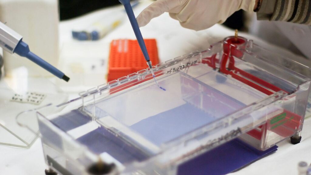
Laboratories use gel separation to confirm sample identity, assess nucleic acid integrity, and verify protein behavior before broader analysis. Reliable results depend on more than a gel and a power source. Chamber geometry, buffer contact, thermal control, and image capture all influence band definition. Careful equipment choices help staff protect limited samples, reduce repeat runs, and compare findings across teaching, research, clinical, and quality settings.
Equipment Selection
Choosing gel electrophoresis equipment begins with sample chemistry, gel format, lane demand, and documentation needs. Horizontal systems work well for DNA and RNA checks. Vertical units enable tighter control of protein separation. Matching trays, combs, power supplies, stains, and imagers keep bands readable, reduce handling, and support dependable interpretation.
Separation Basics
Electrophoresis separates charged molecules as current passes through a buffered gel matrix. Smaller fragments usually migrate farther, while larger material moves more slowly. Gel percentage changes pore size, which affects resolution. Buffer composition influences conductivity and heat. Stable hardware keeps voltage, contact points, and temperature within useful limits during the run.
Chamber Design
A chamber should evenly hold a buffer and keep the electrodes properly aligned. The lid needs secure seating and clear safety markings. Trays should sit flat, because slight movement can skew lanes. Comb slots must form wells with consistent depth. Clear construction helps staff see bubbles, leaks, or loading problems before samples are lost.
Power Control
Power supplies need steady voltage, current, or wattage, depending on the protocol. Small output drift can blur bands or alter migration distance. Timers, readable screens, and protective limits support cleaner operation. Multiple outlets are helpful on busy benches, provided each connected chamber uses compatible settings and sound electrical practice.
Gel Formats
Agarose remains common for nucleic acid work because casting is simple, and concentration can be adjusted. Polyacrylamide provides finer separation for proteins and short fragments. Precast gels reduce preparation variation and save staff time. In-house casting gives teams control over thickness, percentage, and well pattern for specialized assays.
Sample Loading
Good loading technique protects data before the current begins. Wells should be visible, intact, and free from ragged edges. Pipettes, tips, and dye must be compatible with the volume being handled. Overloading can cause streaks, spillover, or distorted lanes. Consistent hand position helps staff compare bands across batches, operators, and benches.
Visualization Tools
After separation, bands must be detected without sacrificing clarity. Blue-light platforms can reduce sample damage when compatible stains are used. Ultraviolet transilluminators still serve many established dye systems. Imaging instruments create records for reports, audits, and peer review. Exposure settings should reveal faint bands while preserving stronger signal boundaries.
Documentation Value
A gel image becomes evidence only when context travels with it. File names, ladder details, run conditions, and exposure settings support traceability. Dedicated capture systems often preserve scale and contrast better than phone images. Consistent records help colleagues compare results across dates, projects, instruments, and separate laboratory areas.
Workflow Fit
Equipment should reflect actual bench activity. Teaching laboratories often need durable chambers, simple controls, and parts that tolerate frequent handling. Research groups may need higher lane capacity, flexible accessories, and imaging options. Regulated spaces often prioritize traceable settings and repeatable records. Practical fit matters more than unused functions.
Maintenance Habits
Routine care protects both instruments and results. Staff should rinse compatible residues, inspect seals, and check electrodes for corrosion. Power leads need tight connections and intact insulation. Imaging surfaces should remain clean, dry, and scratch-free. Brief maintenance logs help teams notice wear before it changes band quality.
Common Errors
Weak gels often stem from avoidable setup errors. Expired buffer, wrong concentration, reversed leads, or damaged combs can reduce definition. Uneven benches may cause liquid to shift during loading. Excess heat can cause bands to curve or lanes to widen. A short pre-run checklist protects samples and preserves staff time.
Purchase Criteria
Before purchase, laboratories should compare chamber dimensions, lane count, gel compatibility, safety features, and service support. Accessory supply matters as much as the main unit. Replacement trays, combs, lids, and leads should be readily available. Clear product data helps teams match equipment with current protocols and expected workload.
Conclusion
Reliable gel analysis depends on equipment that suits the method, specimen type, and laboratory pace. Chambers, power supplies, gels, stains, and imagers function as a connected system. Thoughtful selection reduces variation and makes interpretation more dependable. With careful setup, clear documentation, and routine maintenance, electrophoresis remains a trusted method for daily molecular and protein work.

 Yplostylia Varkonin