Many people hear “eye exam” and think of an eye chart, a glasses prescription, and the familiar question: “Which is better, one or two?” Those steps can be part of the visit, but a comprehensive medical eye exam goes further. It looks at how well a person sees and how healthy the eye structures are.
That matters because some eye diseases can begin before vision changes are obvious. Vision may seem normal while changes are already beginning in the retina, optic nerve, lens, cornea, or blood vessels inside the eye.
Gregory T. Clariday, M.D., from Coastal Eye Associates, explains that people looking for an ophthalmologist in Houston often need more than a routine vision check. Symptoms such as glare, eye pain, floaters, fluctuating blur, headaches, dry eye, or trouble driving at night may call for an exam that looks beyond a prescription.
A Vision Check Is Only the Starting Point
A vision check measures how clearly someone sees at a distance and, when needed, up close. This may include reading letters on an eye chart and testing different lenses to see whether glasses or contacts could sharpen vision. It is useful, but it is only one layer of the exam.
A comprehensive medical eye exam also asks why vision has changed. Blurry vision could come from a simple prescription shift, but it could also be related to cataracts, dry eye, corneal disease, medication effects, diabetes, inflammation, or changes in the retina. Two people may describe the same symptom and need very different next steps.
The medical history is part of the testing process. The doctor may ask about diabetes, high blood pressure, autoimmune disease, migraines, previous eye surgery, steroid use, family history of glaucoma, eye injuries, and current medications. These details help guide what the doctor looks for during the exam.
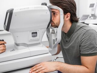
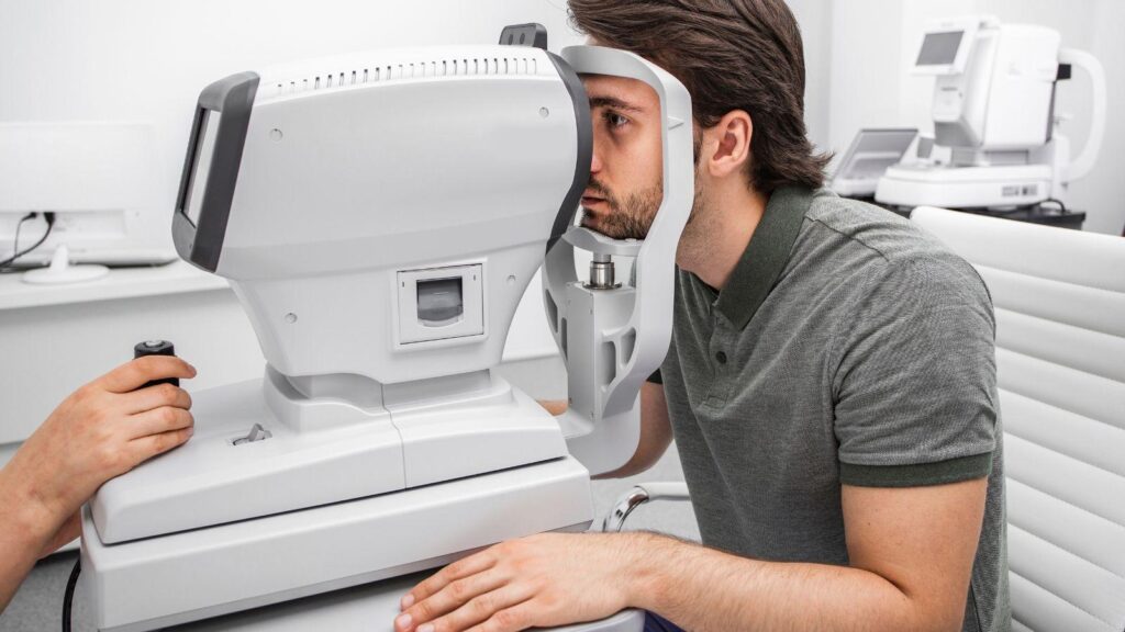
The front of the eye is usually examined with a slit lamp, a microscope with a bright light that gives a magnified view of structures such as the eyelids, cornea, conjunctiva, sclera, and iris. MedlinePlus describes the slit lamp as one of the magnifying devices used during a standard eye exam, and tonometry may also be used to check eye pressure.
That is why a medical exam should not feel like a pass-fail test. Reading the chart is helpful, but it does not answer every question about eye health.
Eye Pressure Testing Helps Screen for Glaucoma Risk
Eye pressure testing, also called tonometry, is one of the exam steps patients often remember. Some people know it as the “air puff” test. Other offices may use different instruments to measure intraocular pressure, which is the pressure inside the eye.
This test is important because eye pressure is one risk factor for glaucoma. Glaucoma is a group of diseases that damage the optic nerve, the nerve that carries visual information from the eye to the brain. The National Eye Institute notes that glaucoma often has no early symptoms and that diagnosis involves a dilated eye exam with visual field testing.
Still, eye pressure alone does not diagnose glaucoma. Some people have higher-than-average pressure and never develop optic nerve damage. Others can develop glaucoma even when their pressure is within a range that might otherwise be considered normal. That is why the doctor may also examine the optic nerve, check peripheral vision, review family history, and sometimes order imaging.
A visual field test may be used when glaucoma is suspected or being monitored. This test checks side vision, where glaucoma-related changes often begin. Patients may not notice early peripheral vision loss because the brain can compensate and the better eye may mask changes in the other eye. By the time a person notices missing side vision in daily life, the disease may already be more advanced.
For people at higher risk, including those with a family history of glaucoma and adults in higher-risk age or demographic groups, regular medical eye exams are especially important. The National Eye Institute identifies higher-risk groups as African Americans over age 40, everyone over age 60, especially Hispanics/Latinos, and people with a family history of glaucoma. The goal is not to make every patient worry about glaucoma. The goal is to find risk early enough to monitor it, treat it, or rule it out.
Retina Evaluation Can Find Problems Before Symptoms Appear
The retina is the light-sensitive tissue lining the back of the eye. It is where light signals are converted into nerve signals that travel to the brain. A retina evaluation helps the doctor look for changes involving blood vessels, the macula, the optic nerve, and the surrounding retinal tissue.
Dilation is often used for this part of the exam. Dilating drops widen the pupil, allowing the doctor to see more of the inside of the eye. The drops can cause temporary light sensitivity and blurred near vision, so patients may want to bring sunglasses and ask whether they should have someone drive them home.
A dilated exam can be especially important for people with diabetes. The CDC advises people with diabetes to get a dilated eye exam at least once a year so an eye doctor can spot problems early. Diabetes can damage the small blood vessels in the retina, leading to diabetic retinopathy. The National Eye Institute explains that eye doctors check for diabetic retinopathy as part of a dilated eye exam and may use additional testing, such as a fluorescein angiogram, when more severe disease or diabetic macular edema is suspected.
Retina evaluation can also help identify signs of macular degeneration, retinal tears, retinal detachment risk, bleeding, inflammation, or changes related to high blood pressure. Some of these issues can cause obvious symptoms, such as flashes, new floaters, dark spots, or distorted central vision. Others may be found before a patient notices anything unusual.
This is one reason medical eye exams can feel more detailed than expected. The doctor is not only asking, “Can you see clearly today?” The larger question is whether the structures responsible for vision look healthy enough to keep working well.
Imaging and Follow-Up Tests Add More Detail When Needed
Not every patient needs every test at every visit. A comprehensive medical eye exam is guided by symptoms, age, health history, risk factors, and what the doctor sees during the exam. When more information is needed, imaging and follow-up testing can add detail.
Optical coherence tomography, often called OCT, creates detailed images that can help doctors evaluate the retina and macula. The National Eye Institute describes OCT as a test that takes pictures of the retina and other parts of the eye, which can help doctors see how much swelling is present in conditions such as macular edema.
In glaucoma care, doctors may also use imaging or visual field testing to track whether the optic nerve or side vision changes over time. Cleveland Clinic describes glaucoma testing as a group of procedures that may include an angle exam, corneal thickness measurement, dilated eye exam, eye pressure check, optic nerve imaging, and visual field testing.
Other tests can add different kinds of information. Color vision testing may be used when color perception is part of the concern. Corneal measurements may help when the doctor needs more information about corneal shape, contact lens concerns, surgical planning, or certain corneal conditions. Tear evaluation or eye-alignment testing may be used when symptoms or exam findings point in those directions.
Follow-up testing does not always mean something serious has been found. Sometimes it means the doctor wants a baseline measurement. Sometimes a finding is borderline and needs to be watched. Sometimes a symptom does not match the first test results, so a more specific test is needed.
This is where a medical eye exam becomes part of long-term care rather than a single appointment. Results can be tracked over time. Small changes in eye pressure, optic nerve appearance, retinal thickness, cataract progression, or visual field results may become more meaningful when compared with previous visits.
For patients who need more than a basic vision check, Coastal Eye Associates provides ophthalmology and optometry care across multiple Houston Bay Area locations, helping connect symptoms with possible causes such as cataracts, glaucoma, retina conditions, or dry eye.
A medical eye exam is not meant to make routine vision care sound complicated. It is meant to give a fuller answer. If the issue is simply a prescription change, that is useful to know. If the exam finds signs of disease early, that information can be even more important. The purpose of these tests is to connect what a patient notices with what the doctor can see inside the eye.