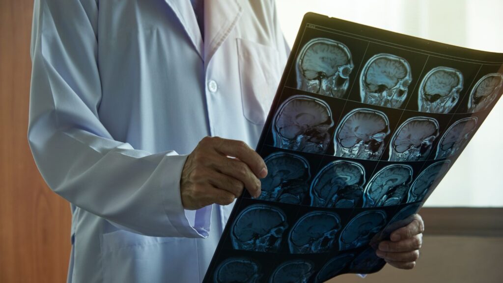
Magnetic Resonance Imaging (MRI) has become one of the most critical tools in modern medicine, offering detailed insights into the human body without the need for invasive procedures. The continuous evolution of MRI technology has particularly transformed the field of neurology, enabling physicians to detect subtle changes in brain structure and function long before clinical symptoms appear. In emergency or diagnostic settings, access to advanced facilities such as the Lumberton Emergency Room plays a crucial role in ensuring timely evaluation and treatment, especially when early neurological signs are present.
Recent advancements in MRI imaging now allow researchers and clinicians to visualize neurodegenerative diseases, such as Alzheimer’s, Parkinson’s, and multiple sclerosis (MS), with unprecedented accuracy. Early detection not only supports faster intervention but also enhances the potential for personalized care and improved patient outcomes.
Detecting Neurodegenerative Diseases Before Symptoms Arise
Traditional neurological diagnosis often relies on behavioral and cognitive assessments, which typically occur only after symptoms manifest. However, neurodegenerative diseases begin silently, with pathological changes developing years before noticeable impairments occur. MRI technology bridges this diagnostic gap by identifying microstructural and biochemical alterations in the brain long before cognitive or motor symptoms appear. In many cases, early access to reliable emergency care services and advanced imaging can make a crucial difference, ensuring faster diagnosis and better treatment planning.
For example:
- Alzheimer’s Disease: Advanced MRI can detect hippocampal atrophy and disruptions in white matter integrity years before memory loss is evident.
- Parkinson’s Disease: Quantitative MRI can identify degeneration in the substantia nigra, a region essential for dopamine production, aiding early therapeutic planning.
- Multiple Sclerosis: MRI remains the gold standard for detecting demyelinating lesions in the central nervous system, helping clinicians initiate treatment early and track disease progression.
These applications are expanding rapidly with the integration of artificial intelligence (AI) and machine learning algorithms, which can analyze vast amounts of imaging data to predict disease risk and progression patterns.
Functional MRI and Brain Connectivity
Functional MRI (fMRI) has revolutionized the understanding of brain activity. Unlike traditional MRI, which captures static images, fMRI measures changes in blood flow that occur when neurons are active. This allows scientists to observe brain networks in real-time, revealing how regions communicate and how those connections deteriorate in neurodegenerative conditions.
In Alzheimer’s disease, for example, fMRI can detect disruptions in the brain’s default mode network, a set of regions responsible for memory and introspection before significant cognitive decline occurs. Similarly, in Parkinson’s disease, functional imaging can identify abnormalities in motor network connectivity, aiding early diagnosis and therapy design.
Diffusion MRI: Mapping White Matter Integrity
Diffusion MRI techniques, such as diffusion tensor imaging (DTI), allow researchers to map the brain’s white matter tracts. These pathways are essential for communication between brain regions, and damage to them is often one of the earliest indicators of neurological disease. DTI metrics like fractional anisotropy (FA) and mean diffusivity (MD) can highlight changes in tissue integrity even before structural atrophy becomes visible.
This capability has opened new possibilities for monitoring disease progression and evaluating treatment effectiveness. In clinical research, DTI is increasingly used as a biomarker for neurodegenerative disease staging, providing valuable insights into the underlying pathology.
Quantitative MRI and Biomarker Discovery
Quantitative MRI (qMRI) takes imaging beyond visualization by assigning numerical values to tissue properties such as T1, T2, and proton density. This standardization allows for consistent comparisons across patients and time points, enhancing the precision of longitudinal studies.
Recent studies show that quantitative MRI can detect iron accumulation, myelin loss, and subtle volumetric changes associated with early-stage neurodegeneration. These quantifiable biomarkers are paving the way for more objective diagnostic criteria and improved monitoring of therapeutic responses.
Artificial Intelligence and Automated Image Analysis
Artificial intelligence (AI) is playing a transformative role in the future of MRI-based diagnostics. Machine learning algorithms can detect complex imaging patterns that might be overlooked by human observers, offering predictive models for early disease detection. Automated image segmentation and pattern recognition are improving diagnostic accuracy and reducing interpretation time, enabling faster and more reliable results.
As datasets continue to grow, AI will be key in integrating imaging data with genetic, biochemical, and clinical information to develop comprehensive models for disease prediction and patient-specific treatment planning.
The Future of MRI in Preventive Neurology
The convergence of advanced MRI techniques, AI analytics, and data integration marks a new era in preventive neurology. Early identification of neurodegenerative processes means that clinicians can intervene sooner, slow disease progression, and improve patients’ quality of life. Moreover, MRI biomarkers are becoming invaluable tools in clinical trials, helping researchers evaluate the effectiveness of emerging therapies with precision.
Integrating Technology and Patient Care
At its core, the advancement of MRI technology underscores the synergy between scientific innovation and patient care. Continuous improvements in imaging precision and data analysis are empowering clinicians to detect neurological changes earlier and design more effective, patient-centered treatment strategies. MRI continues to evolve from a diagnostic tool into a predictive instrument for early disease prevention, reshaping the landscape of modern neurology.
As medical science progresses, platforms like OpenMedScience play an essential role in bridging the gap between research and real-world clinical application. By highlighting breakthroughs in imaging, diagnostics, and medical technology, OpenMedScience fosters collaboration among scientists, healthcare professionals, and innovators driving a deeper understanding of how advancements like MRI can transform patient outcomes and the future of medicine.
Disclaimer
The future of MRI in detecting neurodegenerative diseases is bright and full of promise. With ongoing improvements in resolution, functional imaging, and AI-powered analytics, MRI is not only enhancing early diagnosis but also redefining how medicine approaches prevention and personalized care.


